The Architecture of Human Vision: From Light Waves to Conscious Perception
Human sight is an intricate biological marvel. Every waking second, millions of electromagnetic vibrations pass through living optical elements, converting raw ambient illumination into vivid spatial comprehension within the brain.
1. The Optical Mechanics of the Eye (Cornea, Pupil, and Lens)
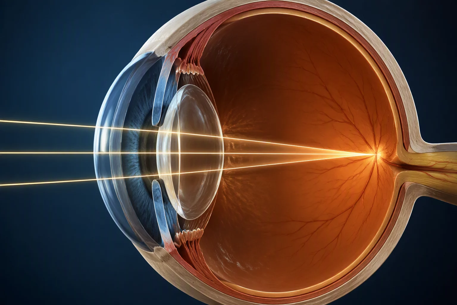
The journey of visual perception begins when light rays reflect off physical surfaces and reach the anterior segment of the human ocular structure. Before neural processing occurs, incoming rays must be precisely bent and focused onto the receptive tissue at the rear of the eye.
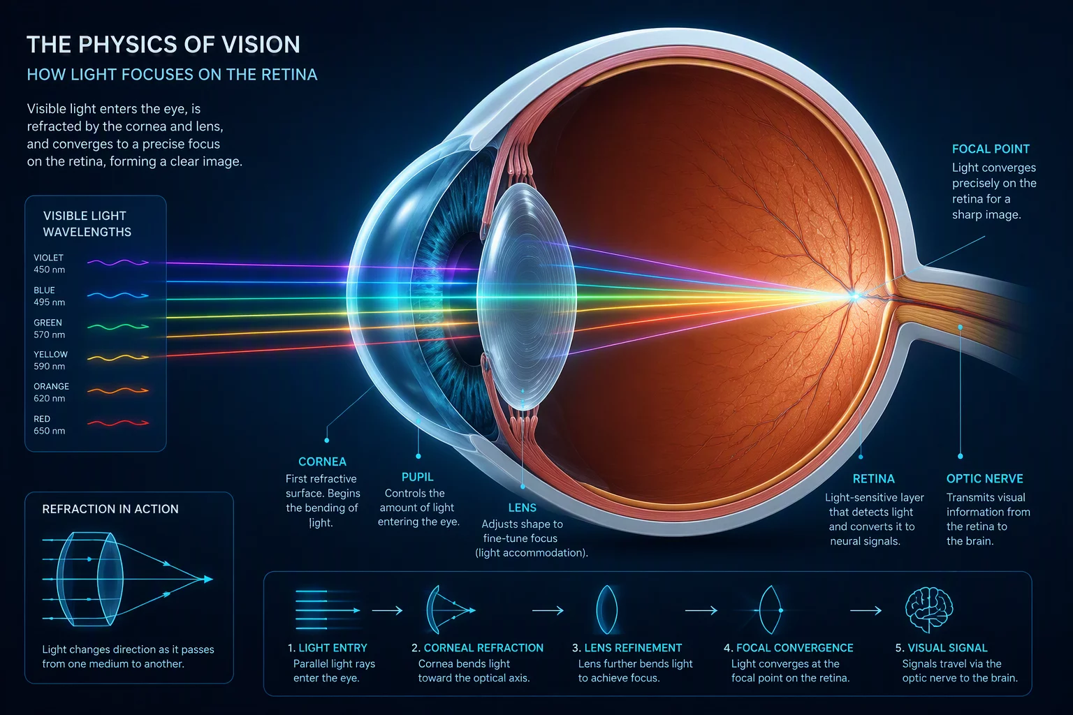
The process depends on three synchronized structural components:
- The Cornea: A clear, dome-shaped outer window responsible for approximately two-thirds of the eye's total refractive optical power. It bends divergent light waves into a converging beam.
- The Pupil and Iris: The dynamic aperture system. The pigmented iris adjusts pupillary diameter via muscular contractions, regulating light admission based on ambient brightness.
- The Crystalline Lens: A flexible, biconvex structure situated directly behind the pupil. Controlled by ciliary bodies, it alters its focal curvature (accommodation) to sharply project near or distant objects onto the retinal plane.
Bio-Physical Phenomenon: Dynamic Focal Adaptation
The human crystalline lens can change optical focus from infinity to twenty centimeters within 350 milliseconds. It continuously recalculates minute refractive discrepancies without conscious mental direction.
2. Photoreception: How Rods and Cones Decode Wavelengths
Once focused photons strike the neural retina, physical electromagnetic energy undergoes transduction into electro-chemical signals. This critical translation is carried out by specialized photoreceptor cells populated across the retinal surface.
The human retina relies on two primary classes of photoreceptors, each engineered for distinct environmental conditions:
- Rods (Scotopic Vision): Numbering over 100 million, rods exhibit extraordinary sensitivity to minimal photon quantities. They excel at detecting motion, contrast, and peripheral silhouettes under low-light conditions but do not distinguish chromatic hues.
- Cones (Photopic Vision): Concentrated predominantly within the fovea centralis, approximately 6 million cone cells operate in well-illuminated environments. They provide high-acuity spatial resolution and tri-chromatic color identification.
Human trichromacy functions through three cone varieties tuned to distinct wavelengths: S-cones (short-wave blue ~420 nm), M-cones (medium-wave green ~530 nm), and L-cones (long-wave red ~560 nm). The brain synthesizes overlapping activation spectra into millions of discernible hues.

3. Neural Pathways: From Retina to the Visual Cortex
Transduced signals do not travel directly to consciousness; they undergo progressive compression and integration through several neurological layers before forming mental imagery.
- Retinal Ganglion Cells: Integrate outputs from bipolar and horizontal cells, bundling over one million axons to form the optic nerve.
- The Optic Chiasm: A pivotal crossroad where nerve fibers from the nasal hemiretinas decussate (cross over), allowing binocular data from both eyes to unite for stereoscopic depth calculation.
- Lateral Geniculate Nucleus (LGN): Located in the thalamus, this relay station organizes visual information into distinct streams for movement, depth, color, and high-frequency details.
- Primary Visual Cortex (V1 / Striate Cortex): Situated in the occipital lobe, V1 breaks down complex scenes into basic orientations, boundaries, angles, and spatial frequencies before broadcasting them to higher association areas (V2, V4, and MT).
Bio-Physical Phenomenon: The Visual Processing Relay
The human occipital lobe processes visual data in parallel streams. Motion is decoded milliseconds before color and form, enabling rapid reflex orientation to sudden shifts in the visual field.
4. Visual Adaptation and Environmental Balance
The visual system constantly stabilizes its output across wildly fluctuating ambient environments. Entering a dim auditorium from bright midday sunlight requires biochemical adjustments known as dark adaptation, driven by the regeneration of rhodopsin pigment molecules inside rod cells.
Likewise, balanced environmental ergonomics—such as diffuse indirect illumination, adequate ambient luminance, and frequent distance gaze shifts—help preserve comfortable visual processing throughout modern daily routines.
Core Insights
- Refraction across the cornea and crystalline lens guarantees precise focal geometry upon the neural retina.
- Photoreceptor division of labor allows high-resolution color discernment alongside acute low-light detection.
- Visual perception is a synthesized reconstruction calculated across multiple specialized cortical regions.
- Lighting harmony and thoughtful everyday visual habits encourage balanced perceptual comfort.